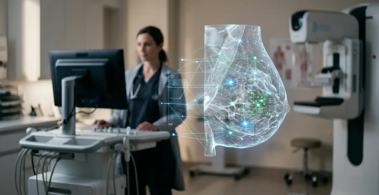
Receiving a callback after a mammogram is incredibly stressful, often due to a ‘false positive’. As a radiologist, I want to reassure you that new, regulated AI tools are now acting as a second pair of expert eyes on your screening. This technology isn’t replacing your doctor; it’s a powerful collaborator that significantly improves accuracy, dramatically reduces false alarms, and helps us give you clearer, more confident answers, faster. The primary goal is to reduce unnecessary anxiety while finding cancers earlier than ever before.
Receiving that phone call is a moment that stops your heart. “We’d like you to come back for a follow-up on your mammogram.” Your mind immediately races. It’s a deeply personal and anxious experience that, as a radiologist, I see women navigate every single day. For many, this callback is the start of a stressful journey of more tests, waiting, and worrying, only to find out it was a ‘false positive’—a benign finding that looked suspicious on the initial scan. While we’ve always preached that early detection is key, we’ve also been acutely aware of the emotional toll of these false alarms.
But what if we could significantly reduce that uncertainty? What if we could bring more confidence to that first reading? This is where the conversation about Artificial Intelligence in medicine moves from a futuristic concept to a present-day reality, especially here in Montreal. The idea of a machine analyzing something as personal as a mammogram might sound cold or impersonal. However, I want to reframe that perspective. Think of it not as a replacement for your doctor, but as an incredibly powerful, diligent, and tireless assistant for your radiology team. Its purpose is to help us separate the ‘signal’ from the ‘noise’ with unprecedented accuracy, reducing the need for those stressful callbacks and giving us, your doctors, more time to focus on what truly matters: you.
This article will walk you through how this collaborative intelligence is changing breast cancer screening. We will explore how these tools are regulated, how they help us reduce overdiagnosis, and why the combination of a human expert and an AI partner leads to the best possible outcome for your health and peace of mind.
Summary: A Radiologist’s Guide to AI in Breast Cancer Screening
- Can a Smartphone Photo Really Diagnose Melanoma Accurately?
- Robots in the Hallway: How Automation Is freeing Up Nurses for Patient Care?
- VR Headsets vs Morphine: Using Tech to Manage Burn Victim Pain?
- How Far Are We From 3D Printing a Kidney for Transplant in Montreal?
- How Blockchain Could Finally Fix the Fax Machine Problem in Healthcare?
- The Risk of Overdiagnosis That Leads to Unnecessary Biopsies
- Pathologist + AI: Why Two Brains Are Better Than One for Cancer Staging?
- How Genomic Sequencing Is Solving Medical Mysteries for Rare Diseases in Kids?
Can a Smartphone Photo Really Diagnose Melanoma Accurately?
You may have seen news about smartphone apps that claim to diagnose skin cancer from a photo. It’s a compelling idea, but it also raises valid questions about accuracy and regulation. In healthcare, especially in Canada, we can’t simply launch an app and hope for the best. Any tool used for diagnosis, including those powered by AI, must undergo rigorous testing and approval. This is to ensure it is not only effective but, more importantly, safe. For example, specific AI software used in breast imaging has to be officially licensed; iCAD’s ProFound AI Risk is licensed by Health Canada, which means it has met stringent standards for clinical use.
This regulatory oversight is what separates a consumer wellness app from a clinical diagnostic tool. The medical community is proceeding with what we call “responsible integration.” As a leading research team noted in a recent PMC review, this involves extensive real-world evaluations, transparent reporting of performance, and continuous monitoring to ensure the technology is fair and safe for everyone. This careful, evidence-based approach is fundamental to building trust. So, while a simple photo app for melanoma isn’t yet a replacement for a dermatologist, the principles of rigorous validation we’re applying in that field are the same ones ensuring the AI used for your mammogram is trustworthy and effective.
Robots in the Hallway: How Automation Is freeing Up Nurses for Patient Care?
Walking through a modern Montreal hospital, you might see robots delivering linens or lab samples. It’s easy to see how this automation frees up staff to focus on patients. In radiology, we’re seeing a similar, though less visible, revolution. The “robot” is the AI software, and its task is to handle a crucial but time-consuming part of our workflow: the initial review of mammograms. A radiologist’s day involves looking at hundreds of images, searching for subtle signs that could indicate a problem. It’s a task that requires immense focus.
By having an AI perform an initial analysis, we can streamline this process significantly. This isn’t about replacing the radiologist but augmenting our ability. The AI can quickly identify and clear the vast majority of clearly normal scans, leaving us more time and cognitive energy to focus on the complex cases or those where the AI has flagged a potential area of concern. The impact is substantial; research from Denmark demonstrates a 33.4% reduction in radiologists’ reading workload when using AI assistance. This reclaimed time is invaluable. It translates into more thorough analysis of difficult scans and, crucially, more time to consult with colleagues and communicate with patients like you.
VR Headsets vs Morphine: Using Tech to Manage Burn Victim Pain?
In other areas of medicine, technology is achieving things that once seemed like science fiction. For instance, some hospitals are now using Virtual Reality (VR) headsets to manage the excruciating pain of burn victims during wound care, sometimes proving as effective as high-dose opioids. This is a powerful example of how innovative thinking can fundamentally improve the patient experience. In breast cancer screening, our “game-changing” technology is AI, and its goal is equally transformative: to catch cancer at its earliest, most treatable stage.
The entire purpose of mammography screening is to find cancer before it has a chance to spread. When we succeed, the impact on a person’s life is profound. The data on this is incredibly motivating for us as clinicians. According to figures highlighted by breast health technology leaders, there is a 99% 5-year relative survival rate when breast cancer is detected early and is still localized within the breast. This single statistic is why we are so passionate about improving our detection tools. AI helps us get closer to this goal by enhancing our ability to spot subtle, early-stage cancers that might be missed by the human eye alone, making this life-saving difference for more women.
How Far Are We From 3D Printing a Kidney for Transplant in Montreal?
The idea of 3D printing a custom organ for transplant, a field where Montreal researchers are very active, represents a futuristic frontier in medicine. It promises a perfect solution to a complex problem. In breast imaging, AI is not a future promise; it’s a “here and now” technology delivering a more immediate, practical solution to the persistent problems of cancer detection. One of the most significant challenges in mammography is balancing the need to find every cancer (sensitivity) with the need to avoid flagging benign issues (specificity). This is precisely where AI is making its biggest impact.
Instead of just talking in concepts, let’s look at the data. A recent large-scale study directly compared traditional screening with AI-assisted screening. The results, as shown in the table below, are compelling for any woman who has worried about a false alarm.
| Method | Cancer Detection Rate | False-Positive Rate |
|---|---|---|
| Traditional Screening | 0.70% | 2.39% |
| AI-Assisted Screening | 0.82% | 1.63% |
What this table shows is the holy grail of screening: we are finding more cancers while simultaneously and significantly lowering the rate of false positives. This isn’t a small adjustment; it’s a fundamental improvement in performance. As a research team studying AI in mammography noted, the integration of AI into breast cancer screening workflows has the potential to not only improve efficiency but also to give patients more accurate and reliable results from the very first scan.
How Blockchain Could Finally Fix the Fax Machine Problem in Healthcare?
It’s a frustrating reality that in an age of instant communication, healthcare can still feel stuck in the past, sometimes literally relying on fax machines. In Quebec, even with advancements like the Dossier santé Québec (DSQ), ensuring different parts of the system talk to each other seamlessly remains a challenge. This isn’t just an inconvenience; it can have real consequences. As a comprehensive review of AI’s role in Quebec healthcare points out, the current system is stretched thin by many issues. These include the risk of overdiagnosis in low-risk groups, the immense workload on radiologists, and inconsistencies in how tests are interpreted across different facilities.
These systemic issues are precisely what AI is poised to address. It’s not a magic bullet for all problems, but it provides a powerful, standardized tool to mitigate some of our biggest hurdles. By providing a consistent, data-driven analysis, AI can help standardize interpretations, reducing the variability between one reader and another. It directly tackles the workload problem, as we’ve seen, and its ability to reduce false positives helps combat the issue of overdiagnosis and the cascade of unnecessary tests that follow. AI is not just a shiny new object; it is a targeted intervention designed to solve long-standing, frustrating problems within our healthcare infrastructure.
The Risk of Overdiagnosis That Leads to Unnecessary Biopsies
The word “overdiagnosis” might sound strange. How can finding something be a bad thing? In screening, it refers to identifying abnormalities that are technically “cancer” but are so slow-growing they would never have caused harm in a person’s lifetime. More commonly, however, the anxiety comes from a “false positive,” where a benign cyst or dense tissue is flagged as suspicious, leading to a callback. This callback is what triggers a cascade of more imaging, anxiety, and often, an unnecessary biopsy. This is the single biggest downside of screening, and it’s the area where I, as a radiologist, see the most immediate benefit of AI.
AI is exceptionally good at pattern recognition, at learning what is a truly suspicious finding versus what is just unusual but benign breast tissue. It acts as a sophisticated filter, helping us separate the crucial signals from the background noise. The results are not just theoretical. A major Danish study found that AI-assisted screening achieved a 20.5% decrease in the recall rate. That means one in five women who would have been called back for a stressful follow-up no longer have to be. As Dr. Richard L. Wahl of Washington University School of Medicine eloquently stated, this is about more than just efficiency. In his words:
False positives are when you call a patient back for additional testing, and it turns out to be benign. That causes a lot of unnecessary anxiety for patients and consumes medical resources.
– Richard L. Wahl, MD, Washington University School of Medicine
This directly addresses the heart of the problem: reducing the emotional and financial cost of false alarms, which is a massive step forward for patient-centered care.
Pathologist + AI: Why Two Brains Are Better Than One for Cancer Staging?
When a biopsy is performed, a pathologist examines the tissue under a microscope to confirm a diagnosis and determine its characteristics—a process called staging. Just as in radiology, AI is becoming a powerful collaborator for pathologists. The core principle is “collaborative intelligence”—the idea that a human expert paired with an AI partner is far better than either one working alone. The AI can analyze the entire tissue sample at a microscopic level, flagging areas of interest that the pathologist can then focus on with their expert judgment. It’s the ultimate “second read.”
The performance of these systems is remarkable, not as a replacement, but as an enhancement. One study showed a leading AI system was better at distinguishing cancer on slides than the average of its human counterparts, but the real magic happens when they work together. However, we must be realistic. Integrating these tools into the complex clinical environments at hospitals affiliated with McGill or UdeM is a challenge. We have to address issues of model generalizability (making sure an AI trained in one hospital works well in another) and navigate the “black-box” problem—understanding *why* the AI made a certain recommendation. This ensures we are always in control and using the technology responsibly.
Your Checklist for Discussing AI with Your Doctor
- Ask the question: Does this hospital or clinic use AI-assistance in its mammogram reading process?
- Clarify its role: Can you explain how the technology helps you as the radiologist make a decision?
- Inquire about the process: If AI flags something, what are the next steps? Is it always reviewed by a human expert? (The answer should always be yes).
- Discuss your concerns: Feel free to share any anxieties you have about the technology. Your healthcare provider is there to help you understand.
- Request information: Ask if there are any patient-friendly brochures or web pages that explain their use of the technology.
This approach ensures we leverage the power of AI while upholding the highest standards of medical practice and patient trust.
Key Takeaways
- AI in mammography acts as a “second reader,” helping radiologists find more cancers while significantly reducing false-positive callbacks.
- This technology is strictly regulated by bodies like Health Canada to ensure safety and effectiveness before being used in clinics in Montreal and across the country.
- The main patient benefit is a reduction in “scanxiety”—the stress and worry caused by unnecessary follow-up tests and biopsies.
How Genomic Sequencing Is Solving Medical Mysteries for Rare Diseases in Kids?
Just as genomic sequencing is unlocking personalized treatments by reading our genetic code, AI is helping us read medical images with a new level of understanding. However, with any powerful new technology, we must also be aware of its limitations and potential pitfalls. An AI system is only as good as the data it’s trained on. If the training data is not diverse, the AI can develop blind spots or biases. This is a topic the entire medical and tech community takes extremely seriously.
As one leading AI researcher, Meredith Broussard, has pointed out, pre-existing biases in the world can easily get embedded in the systems we build. For example, if an AI is primarily trained on images from one demographic group, it may be less accurate for others. As she states, this is a critical challenge:
There is a lot of bias in AI because there’s a lot of bias in the world, and that pre-existing bias gets embedded in AI systems.
– Meredith Broussard, New York University AI researcher
Acknowledging this is the first step to solving it. Here in Montreal and across the global research community, a huge amount of effort is focused on creating diverse, representative datasets and developing techniques to audit AI for fairness. Our ethical obligation is to ensure that these powerful tools benefit everyone equally. It’s a continuous process of improvement, and this transparency about the challenges is just as important as celebrating the successes.
The journey of integrating AI into breast cancer screening is about one thing: improving your care. It’s about giving you and your healthcare team more confidence in your results, reducing the emotional burden of uncertainty, and focusing our collective expertise on finding and treating cancer earlier and more effectively than ever before. The next time you have a mammogram, know that your radiology team in Montreal is equipped with the best of both human and artificial intelligence, all working together for you.