You’ve been sent for a specific MRI, and it’s confusing because the hospital is far or the wait is long; the key is that your doctor hasn’t just chosen a ‘stronger’ machine, but the right ‘diagnostic lens’ for your unique brain scan.
- A 3T MRI offers higher-resolution images, crucial for detecting subtle neurological conditions, but isn’t always necessary.
- Montreal’s healthcare system presents a choice: a long but covered (RAMQ) wait for a public hospital scan, or a fast (24-48 hours) but expensive ($900+) private clinic appointment.
Recommendation: Understand the clinical reason behind your referral and use this guide to navigate your options in Montreal, whether you choose the public or private path.
Holding a doctor’s referral for an MRI in Montreal can feel like the start of a complicated journey. You might be wondering, “Why this specific hospital? Why a 3T machine? And why is the wait so long?” It’s a common point of confusion. Many people assume that a 3 Tesla (3T) MRI is simply “better” or “stronger” than a 1.5 Tesla (1.5T) machine, and that they should always demand the most powerful option available.
As an MRI technologist, I can tell you the reality is more nuanced. The choice between a 1.5T and a 3T machine isn’t about raw power; it’s about choosing the right tool for the job. Think of it like a professional photographer selecting a camera lens. For a standard portrait, a reliable workhorse lens is perfect. But to capture the intricate details on a butterfly’s wing, a specialized macro lens is required. Your neurologist is making a similar choice for your brain.
This guide is designed to demystify that choice. We’ll break down what these numbers—1.5T and 3T—actually mean for your scan’s image quality. We’ll explore the specific situations where a high-definition scan is non-negotiable and what that means for navigating the waitlists and costs within Montreal’s public and private healthcare systems. The goal is to replace your uncertainty with understanding, empowering you to feel confident in the diagnostic path your doctor has chosen.
In this guide, we will walk through the key differences between these technologies and what they mean for you as a patient in the Montreal area. The following sections break down everything from image quality to wait times, helping you understand your referral.
Summary: A Patient’s Guide to 1.5T and 3T Brain MRIs in Montreal
- Why the Louder MRI Machine Often Gives Better Pictures?
- Can You Get an MRI If You Have a Titanium Knee Replacement?
- Seeing Thought: How fMRI Maps Brain Activity Before Neurosurgery?
- Why the Wait List for High-Definition MRI Is Twice as Long?
- Gadolinium Retention: Is It a Real Risk for Repeat MRI Patients?
- How to Find a Private MRI Appointment in Montreal in Under 48 Hours?
- Public Hospital vs Private Clinic: Which Path Suits Your Urgency?
- Why an MRI of the Central Nervous System Is More Detailed Than a CT Scan?
Why the Louder MRI Machine Often Gives Better Pictures?
The first thing many patients notice about a 3T MRI is that it’s louder than a 1.5T. That increased noise is a side effect of the stronger hardware working harder to produce a more detailed image. The “T” stands for Tesla, a unit of magnetic field strength. A 3T machine has a magnet twice as strong as a 1.5T, and this is the key to its superior image quality. A stronger magnet allows us to get a stronger signal from your body’s tissues. We call this the Signal-to-Noise Ratio (SNR). Think of it as trying to listen to a quiet voice in a crowded, noisy room. The 3T machine essentially makes that voice much louder and clearer, so we can distinguish it from the background noise.
For a complex brain scan, this is critical. A higher SNR means we can create images with higher resolution, allowing radiologists to see smaller details, like tiny lesions in multiple sclerosis, early-stage tumors, or subtle changes in brain structure related to epilepsy or dementia. In fact, a 3T MRI scanner delivers a 30-85% practical gain in signal-to-noise ratio compared to its 1.5T counterpart. This isn’t just a technicality; it can be the difference between detecting a condition and missing it. This is why for specific neurological questions, your doctor insists on a 3T scan—they’ve chosen the high-detail “macro lens” to get the clearest possible picture of what’s happening inside your brain.
This enhanced capability is backed by extensive research. For instance, in a 2015 study for the Alzheimer’s Disease Neuroimaging Initiative, researchers made it clear:
3T showed a superior signal-to-noise ratio and detected atrophy with greater effect size compared with 1.5T.
– Alzheimer’s Disease Neuroimaging Initiative researchers, Journal of Neuroradiology
Can You Get an MRI If You Have a Titanium Knee Replacement?
This is one of the most common questions we get from patients, and it’s an important one. The short answer is: almost always, yes. For decades, the presence of metal in the body was a major contraindication for MRI due to the powerful magnet. However, modern medicine has evolved. Most surgical implants used today, especially those for joint replacements like knees and hips, are made from titanium or other non-ferromagnetic materials. These materials are not attracted to the magnet and are considered “MR Safe.”
While the implant itself is safe, it can sometimes cause image distortion, known as an “artifact,” in the area immediately surrounding it. This is like a bright glare in a photograph. Technologists are trained to use special software sequences to minimize these artifacts. For a brain scan, a titanium knee replacement is very far from the area being imaged, so the risk of distortion is practically zero. It’s more of a consideration for scans of the pelvis or legs. The most crucial thing is full disclosure. You must inform the clinic and the technologist of all implants, surgical clips, or any other metal in your body, no matter how small or how long ago it was placed.
Your Pre-Scan Safety Checklist for Implants in Quebec
- Declare all metal implants, including titanium replacements, when booking and on the safety questionnaire.
- Bring your implant card, provided by your surgeon after the operation, to your MRI appointment.
- The technologist will review your card and questionnaire to confirm the implant is compatible with the MRI’s field strength.
- If you have multiple implants (e.g., dental work, surgical clips), list every single one.
- Follow the technologist’s positioning instructions carefully, as they are designed to ensure your safety and optimize image quality.
Seeing Thought: How fMRI Maps Brain Activity Before Neurosurgery?
Beyond static images of the brain’s structure, MRI technology can do something that feels like science fiction: it can watch the brain at work. This is called functional MRI (fMRI). Instead of just taking a detailed anatomical picture, fMRI measures changes in blood flow to different parts of the brain. When a brain area is more active, it consumes more oxygen, and the fMRI machine can detect the corresponding increase in oxygenated blood. This allows neuroscientists and surgeons to create a map of a patient’s brain, showing which areas are responsible for critical functions like language, memory, and movement.
This technology is indispensable for pre-surgical planning, especially in complex neurosurgery. If a surgeon needs to remove a brain tumor, they must know the exact location of these critical functional areas to avoid them. Before the surgery, the patient will perform tasks while in the MRI scanner—like tapping their fingers or answering questions—and the fMRI map shows the surgeon the precise “geography” of that individual’s brain. This personalized map helps them plan the safest possible route to remove the tumor while preserving the patient’s quality of life. In Montreal, this advanced work is happening at world-class facilities. For example, the McConnell Brain Imaging Centre at McGill University operates both 3T and highly advanced 7T MRI systems specifically for this kind of detailed brain mapping research.
The ability to map out these functional pathways is a perfect example of why a high-field 3T magnet is essential. The blood flow changes that fMRI detects are incredibly subtle. The superior Signal-to-Noise Ratio of a 3T machine is necessary to capture these faint signals with enough clarity to create a reliable and accurate map for a surgeon to use in the operating room.
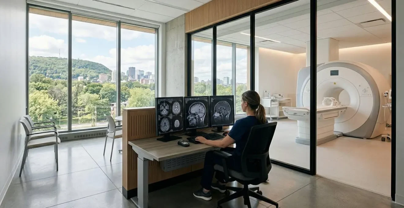
Why the Wait List for High-Definition MRI Is Twice as Long?
If your doctor has ordered a 3T MRI, you’ve likely discovered a frustrating reality of the Montreal healthcare system: the wait can be significantly longer than for a standard 1.5T scan. This comes down to a simple issue of supply and demand. There are far fewer 3T machines in Quebec’s public hospital system. These high-definition scanners are expensive to purchase and maintain, and they are typically reserved for complex cases and advanced research at major university hospitals like the CHUM or the Montreal Neurological Institute.
This scarcity creates a bottleneck. While a 1.5T MRI might have a wait time of a few months, the queue for a specialized 3T scan can easily be double that. According to the Fraser Institute’s 2024 report, patients face a median wait of 16.2 weeks for an MRI across Canada, and this figure is often longer for specialized scans in Quebec. This situation is further complicated by operational issues. A La Presse investigation reported by Global News highlighted that MRI machines in public hospitals are often underutilized, sometimes running for only 8 hours a day instead of the intended 16, while thousands of patients remain on waitlists. This operational inefficiency directly contributes to the long delays you may be experiencing.
This is where the public versus private system dynamic becomes most apparent for patients. The long public wait time is the primary reason many people with the financial means or private insurance opt for a private clinic, where a 3T scan can often be booked within days.
| Service Type | Wait Time | Cost | 3T Availability |
|---|---|---|---|
| Public (RAMQ) | 3-6 months | Covered | Limited, prioritized |
| Private Clinic | 24-48 hours | $900-1500 CAD | Widely available |
Gadolinium Retention: Is It a Real Risk for Repeat MRI Patients?
For some brain scans, your doctor may request an MRI “with contrast.” This means that during the procedure, a technologist will inject a contrast agent—most commonly one containing a rare earth metal called gadolinium—into your bloodstream via an IV. Gadolinium-Based Contrast Agents (GBCAs) make certain tissues, abnormalities, and blood vessels stand out more clearly on the images, providing critical diagnostic information that might be invisible on a non-contrast scan. However, you may have seen news stories about gadolinium retention in the body, leading to patient anxiety.
It’s important to separate the history from the current reality. The concerns originally arose from older types of GBCAs (known as “linear” agents) which were linked to a rare but serious condition called Nephrogenic Systemic Fibrosis (NSF) in patients with severe kidney disease. In response, the medical community has almost universally shifted to using newer, much safer agents called “macrocyclic” GBCAs. These agents have a structure that holds the gadolinium ion much more securely, allowing it to be filtered out of the body by the kidneys much more efficiently, dramatically reducing the risk of retention.
Case Focus: The Safety Profile of Modern Contrast Agents
The safety of modern contrast is not just theoretical. In large-scale studies evaluating the new generation of macrocyclic GBCAs, the results have been overwhelmingly positive. These studies, involving thousands of patients, including those with severe kidney disease who are at the highest risk, have shown that modern agents have an excellent safety profile. In fact, many of these extensive reviews have reported zero confirmed cases of NSF when using macrocyclic agents, confirming their stability and the body’s ability to excrete them effectively. This data provides strong evidence that the benefits of obtaining crucial diagnostic information from a contrast-enhanced MRI far outweigh the now-minimal risks associated with modern agents.
For any patient, the decision to use contrast is based on a careful risk-benefit analysis by your radiologist and referring physician. If they have ordered a scan with contrast, it is because they believe the potential diagnostic information is essential for your care. If you have concerns, especially if you have a history of kidney problems, you should absolutely discuss them with your doctor. However, you can be reassured that the agents used in Montreal clinics and hospitals today are the safest available.
How to Find a Private MRI Appointment in Montreal in Under 48 Hours?
If the public wait time is not a viable option for you due to medical urgency or personal reasons, the private system in Montreal offers a rapid alternative. Securing a private MRI appointment is a straightforward process, but it requires you to be proactive. The key advantage is speed: you can often get a scan, including a complex 3T brain MRI, within 24 to 48 hours. The main trade-off, of course, is the cost, which is paid out-of-pocket or sometimes covered by private insurance plans.
The first and most important step is to have a valid requisition from a licensed physician in Canada. Private clinics cannot perform an MRI without a doctor’s referral. Once you have your requisition, you can start contacting the major private imaging providers in the Montreal area. It’s wise to call a few to compare prices and availability for the specific scan you need. Be sure to specify that you require a brain MRI and whether it needs to be 1.5T or 3T, as indicated on your requisition, as not all private clinics have 3T machines. Prices for a private brain MRI in Montreal typically range from $900 to $1,500 CAD.
Here is a practical, step-by-step guide to navigating the process:
- Obtain a Requisition: This is non-negotiable. You must have a referral form from a licensed physician.
- Contact Providers: Call major Montreal providers like Imagix (Biron), Medvue, VM Med, or Radimed to inquire about 3T brain MRI appointments.
- Compare Prices: Ask for the exact cost of the procedure. Prices can vary between clinics.
- Verify 3T Availability: Explicitly confirm they have a 3T scanner and availability if that is what your doctor has ordered.
- Check Insurance Coverage: Contact your private insurance provider (e.g., Sun Life, Manulife, Desjardins) to see if any portion of the cost is covered under your plan.
- Plan for Results: Confirm that the radiologist’s report will be sent directly to your referring physician and that you will receive a CD with the scan images, which is essential for your specialist or surgeon.
Public Hospital vs Private Clinic: Which Path Suits Your Urgency?
Choosing between the public (RAMQ-covered) and private systems for your MRI in Montreal is a significant decision that balances time, cost, and personal circumstances. There is no single “right” answer; the best path depends entirely on your individual situation. The primary driver for most people is urgency. If your doctor needs a rapid diagnosis to rule out a serious condition or to guide an immediate treatment plan, the weeks or months of waiting in the public system may not be medically advisable. In this case, the private system’s ability to provide a scan within a day or two becomes invaluable.
Conversely, if the MRI is for a non-urgent follow-up, monitoring a stable chronic condition, or if your budget is a primary concern, the public system is the logical choice. The scan will be fully covered by RAMQ, and while the wait is long, the quality of the imaging and the expertise of the radiologists are excellent. It is a common misconception that the quality of care is inferior in the public system. The main difference is not quality, but access and timeliness. A crucial point of reassurance for patients is that the standards are uniformly high across both sectors. As Quebec’s medical regulations state:
Radiologists in both public and private sectors in Quebec are certified by the Collège des médecins.
– Quebec healthcare system documentation
This means that no matter where you get your scan, the expert reading your images is held to the same high professional standard. To help you decide, consider the following factors:
| Decision Factor | Choose Public (RAMQ) | Choose Private |
|---|---|---|
| Medical Urgency | Non-urgent follow-up | Rapid diagnosis needed |
| Financial Situation | Limited budget | Can afford $900-1500 |
| Time Flexibility | Can wait 3-6 months | Need results within days |
| Insurance Coverage | RAMQ only | Private insurance available |
| Specialist Requirements | Standard imaging sufficient | 3T specifically requested |
Key Takeaways
- A 3T MRI isn’t just ‘stronger’; it’s a high-detail ‘diagnostic lens’ with better Signal-to-Noise Ratio, essential for many complex brain scans.
- Navigating Montreal’s system means choosing between a long but covered wait in the public system (RAMQ) or a fast but costly ($900+) private option.
- Safety is paramount: always declare all implants, but know that modern materials like titanium are usually MRI-compatible and modern contrast agents are very safe.
Why an MRI of the Central Nervous System Is More Detailed Than a CT Scan?
Your doctor has referred you for an MRI, but you may have had a CT (Computed Tomography) scan in the past and are wondering what the difference is. While both are powerful imaging tools, when it comes to the brain and central nervous system, MRI is the undisputed champion of detail. The fundamental difference lies in what they “see.” A CT scan is essentially a sophisticated, 3D X-ray. It is excellent at showing dense structures like bone and is very fast, making it the tool of choice for emergencies like head trauma or suspected stroke where speed is critical.
However, an MRI does not use ionizing radiation. Instead, it uses a powerful magnetic field and radio waves to get signals from the water molecules in your body. The brain is about 75% water, and MRI is exquisitely sensitive to the different amounts of water in various types of soft tissue. This allows it to produce images of the brain and spinal cord with stunning contrast and detail that a CT scan could never achieve. It can clearly differentiate between grey matter, white matter, cerebrospinal fluid, and nerves. Think of a CT scan as a high-quality black-and-white photograph—very clear, but limited in its palette. An MRI, in this analogy, is a full-colour, high-resolution photo, revealing a world of subtle shades and textures.
This ability to visualize soft tissue in such detail is why MRI is the gold standard for diagnosing conditions like multiple sclerosis, brain tumors, spinal cord injuries, and the subtle brain changes associated with neurodegenerative diseases. It allows your doctor to see not just the structure, but the health and integrity of the brain tissue itself. The choice to order an MRI over a CT is a deliberate one, made because your doctor needs the unparalleled level of detail that only the “full-colour” view of an MRI can provide.
Armed with this knowledge about the technology and the system, you can now have a more informed conversation with your doctor and feel confident in the specific imaging plan they’ve prescribed for your health.