For a Montreal woman with dense breasts, the choice between a free 2D mammogram and a paid 3D one isn’t about technology—it’s a strategic decision about managing uncertainty and hidden costs.
- 3D mammography significantly reduces anxiety-inducing callbacks by pinpointing issues that look suspicious on a flat 2D image.
- The upfront private cost can be offset by avoiding the financial and emotional toll of false positives, such as lost work days and follow-up fees.
Recommendation: View the 3D mammogram not as an expense, but as a calculated investment in your diagnostic clarity and peace of mind.
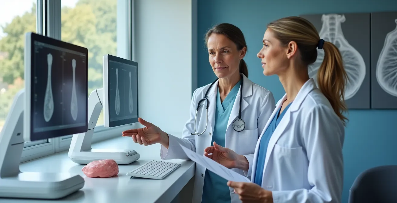
For many women in Montreal, the annual mammogram is a routine part of their healthcare. The Quebec Breast Cancer Screening Program (PQDCS) provides standard 2D mammograms, covered by RAMQ. But for a significant portion of the population, specifically women with dense breast tissue, this routine check-up comes with a cloud of uncertainty. Dense tissue can appear white on a mammogram, the same color as potential tumors, creating a « camouflaging » effect that makes standard 2D imaging less effective and increases the chance of a dreaded callback for more tests.
This is where 3D mammography, or tomosynthesis, enters the picture. It’s offered in private Montreal clinics for a fee, promising a clearer, more detailed view. The common advice is simply that « 3D is better, » but this overlooks the real question a woman is asking herself: « Is it worth several hundred dollars of my own money? » The debate isn’t just about technical specifications; it’s a complex cost-benefit analysis involving money, time, and most importantly, anxiety.
This article reframes the decision. We will move beyond the sticker price to analyze the concept of diagnostic clarity as a personal investment. Instead of just comparing technologies, we will dissect the hidden costs of uncertainty within the public system versus the upfront cost of certainty in the private one. The true value of 3D mammography for a woman with dense breasts in Montreal may not be found on the clinic’s price list, but in the potential to buy peace of mind.
Throughout this guide, we’ll explore this decision from multiple angles, using analogies from other medical fields to clarify the stakes and providing you with the tools to navigate this choice with confidence. We’ll delve into the financial, emotional, and practical implications to help you determine if this advanced imaging is the right strategic investment for your health.
Summary: Deciphering the Value of 3D Mammography in Montreal
- Why Is a CBCT Scan Essential Before Getting Dental Implants?
- Virtual vs Optical Colonoscopy: Which Is Less Invasive for Screening?
- CT Scan Radiation: How Much Is Too Much Within One Year?
- The « Incidentaloma » Trap: Finding Benign Lumps That Cause Anxiety
- How to Breathe During a 3D Scan to Avoid Blurry Images?
- The « Shadow » on the Lung That Turns Out to Be Scar Tissue
- Root Canal ($900) or Extraction ($200): Which Is the Better Long-Term Investment?
- 1.5T vs 3T MRI: Which Machine Should You Choose for Brain Imaging?
Why Is a CBCT Scan Essential Before Getting Dental Implants?
In dentistry, no surgeon would place an expensive implant without a Cone Beam CT (CBCT) scan. A standard 2D X-ray is simply not enough; it doesn’t provide the depth and precision needed to map bone density and nerve pathways. The CBCT is a non-negotiable upfront cost to ensure a successful long-term outcome and avoid catastrophic failure. This principle provides a powerful analogy for breast imaging. For women with dense breasts, a 2D mammogram can be like that flat dental X-ray—it shows the general landscape but lacks the depth to reveal what’s hidden within. Opting for 3D mammography is akin to choosing the CBCT: it’s an investment in seeing the complete picture before making critical decisions.
Of course, this investment has a clear price tag in Montreal’s private healthcare market. The standard 2D mammogram is covered by RAMQ, making it free at the point of service. However, advanced imaging comes at a cost, as it falls outside public coverage.
This table outlines the typical costs for these services in private Montreal clinics, highlighting the financial barrier that is the first part of the patient’s decision-making process.
| Imaging Type | Average Cost (CAD) | Coverage | Detection Improvement |
|---|---|---|---|
| 3D Mammography | $230-350 | Not covered by RAMQ | 41% more invasive cancers |
| CBCT Dental Scan | $300-400 | Not covered by RAMQ | Essential for implant planning |
| 2D Mammography | Free | Covered by RAMQ | Standard detection rate |
Your Action Plan: Questions to Ask Your Montreal Healthcare Provider
- Assess Necessity: Ask « Is this advanced technology strictly necessary for my specific case and breast density? »
- Understand Risks: Inquire, « What are the specific risks of proceeding with only the standard RAMQ-covered option for me? »
- Confirm Costs: Get a clear answer: « How much will the private scan cost at your facility, including all fees? »
- Request Analysis: Ask your doctor, « Can you provide a personalized cost-benefit analysis for my situation, considering my risk factors? »
- Explore Options: Don’t hesitate to ask, « Are there any payment plans or financial assistance available for these private imaging services? »
Facing this direct cost is only the first step. The true evaluation involves weighing this expense against the less tangible, but equally significant, costs of uncertainty and anxiety.
Virtual vs Optical Colonoscopy: Which Is Less Invasive for Screening?
The term « invasive » often brings to mind physical discomfort. In colon cancer screening, a virtual colonoscopy (CT scan) is less physically invasive than a traditional optical one. However, in the context of breast screening, the most invasive part is often not the procedure itself, but the psychological toll of a « false positive. » This is the phone call informing you that your mammogram showed something suspicious and you need to come back for more tests. For a woman with dense breasts, this is a common scenario with 2D imaging. The days or weeks of waiting for follow-up appointments are filled with intense anxiety.
This is where the concept of « anxiety arbitrage » comes into play. By paying for a 3D mammogram, you are investing in a technology that dramatically reduces these false alarms. The 3D view allows the radiologist to see « through » the dense tissue, often confirming immediately that a suspicious shadow is just overlapping tissue, not a lump. As Dr. Majidi from the Froedtert & MCW health network noted in a study, the value is clear:
The 3D mammogram can often prevent that short-term anxiety by reducing callbacks by up to 40% compared to 2D alone.
– Dr. Majidi, Froedtert & MCW health network study
This reduction is a significant benefit. It’s a tangible return on investment measured in peace of mind. Furthermore, the importance of participating in screening cannot be overstated. Organized programs are proven lifesavers. Pan-Canadian data shows up to a 40% lower breast cancer mortality for participants in regular screening programs. Ensuring that the screening experience is as stress-free as possible is key to maintaining this participation. If the fear of a false-positive callback deters a woman from her next screening, the consequences can be severe.
Therefore, the cost of a 3D mammogram can be seen as an insurance premium against weeks of debilitating worry, a factor that is often overlooked in a simple cost comparison.
CT Scan Radiation: How Much Is Too Much Within One Year?
A common and valid concern with any medical imaging is radiation exposure. It’s natural to wonder if the enhanced clarity of 3D mammography comes with a dangerous trade-off. While it’s true that tomosynthesis involves a slightly higher dose than a traditional 2D mammogram, it’s crucial to put this dose into perspective. The combined radiation from a modern 3D mammogram (which includes a synthetically generated 2D image) is extremely low.
According to guidelines from the American College of Radiology, the dose is minimal. For instance, the total dose for a comprehensive 3D scan is about 0.5 millisieverts (mSv) for a synthetic 2D and 3D mammogram combined. To contextualize this, this is less than the background radiation a person in Canada is exposed to from natural sources over a couple of months, and comparable to the dose received during a short flight. The additional dose from a 3D scan is considered negligible by radiologists when weighed against the massive benefit of improved detection in dense breasts.

All radiology clinics in Quebec, both public and private, operate under the ALARA principle— »As Low As Reasonably Achievable. » This means they are legally obligated to use the minimum radiation dose necessary to obtain a high-quality diagnostic image. The equipment is meticulously calibrated and a patient’s exposure is carefully managed. The slight increase with 3D technology is well within established international safety limits and is justified by the significant reduction in callbacks and the increased cancer detection rate.
Ultimately, the risk from the tiny additional radiation dose of a single 3D mammogram is theoretical and minuscule compared to the very real risk of a delayed cancer diagnosis in dense breast tissue.
The « Incidentaloma » Trap: Finding Benign Lumps That Cause Anxiety
While 3D mammography is superior at finding cancers hidden in dense tissue, its high sensitivity comes with a double-edged sword: the « incidentaloma. » This is the medical term for a finding discovered incidentally that is often benign and clinically insignificant, but which can trigger a cascade of further tests, biopsies, and profound anxiety. Tomosynthesis, by its very nature, sees more. It will inevitably detect more small, benign cysts, fibroadenomas, and other harmless lumps that a 2D mammogram might have missed. This is a crucial nuance to understand. The goal is not just to find *more* things, but to find the *right* things.
This is particularly relevant for the Canadian population, where over 40% of women aged 40-74 have dense breasts, a group for whom this technology is most beneficial but also most likely to yield incidental findings. The challenge for the radiologist is to correctly interpret these findings without subjecting the patient to unnecessary follow-up procedures.
Case Study: The Complexities of High Sensitivity
A study at a Canadian screening site highlighted this complexity. It found that while 3D mammography did reduce false positive callbacks caused by simple tissue overlap, its higher sensitivity led to the detection of more small, clinically insignificant findings. A key finding from the study, published in the Canadian Association of Radiologists Journal, showed that mammography’s ability to detect cancer (its sensitivity) varies dramatically with breast density. It can be as high as 93% in fatty breasts (Category A) but drops to as low as 57% in extremely dense breasts (Category D). This demonstrates that while 3D helps, managing the incidental findings it generates in dense breast populations requires significant expertise to avoid over-investigation.
This doesn’t invalidate the value of 3D mammography. On the contrary, it underscores the importance of having the scan read by an experienced breast radiologist who can confidently dismiss benign findings and prevent the « incidentaloma trap, » ensuring that the technology’s power is used for clarity, not for creating new anxieties.
The solution to this trap is not less technology, but more expertise in interpreting its results, which is a hallmark of specialized private clinics.
How to Breathe During a 3D Scan to Avoid Blurry Images?
The advanced technology of a 3D mammogram machine is only half of the equation for achieving diagnostic clarity. The other half is you, the patient. Motion during the scan, even from breathing, can cause blurring on the images, potentially obscuring details and reducing the effectiveness of the exam. While the breast is compressed, the X-ray tube moves in an arc over it, taking multiple low-dose images. Holding perfectly still during these few seconds is critical.
Many patients, understandably anxious, tend to hold their breath deeply or tense their shoulders, which can paradoxically increase body movement. A technologist will guide you, but knowing the proper technique beforehand can empower you and contribute to a better-quality scan. The goal is to be relaxed and still. The compression itself is no different or more painful than a 2D mammogram; it is firm but should not be unbearable. You are always in control and can communicate with the technologist at any time.

Mastering a simple breathing technique can make a significant difference. Here’s a sequence recommended by breast imaging experts:
- Prepare before positioning: As the technologist positions you, take a slow, deep breath in and exhale completely to relax your body.
- The crucial hold: When the technologist says, « Hold your breath, » take a normal, gentle breath in (not a deep gasp) and hold it.
- Stay relaxed: While holding for the 10-15 seconds of the scan, focus on keeping your shoulders down and relaxed, not tensed up towards your ears.
- Communicate: You are in control. If you feel you can’t hold your breath any longer or feel dizzy, signal or tell the technologist immediately. They can stop and restart.
By being an active participant in your exam, you directly contribute to the quality of the images. A sharp, clear image is the foundation of an accurate diagnosis and is the first step in avoiding the need for repeat scans.
This small effort on your part helps maximize the return on your investment in advanced imaging, ensuring the radiologist has the best possible information to work with.
The « Shadow » on the Lung That Turns Out to Be Scar Tissue
In chest X-rays, a « shadow on the lung » is a terrifying phrase for any patient. Yet, frequently, it turns out to be nothing more than old scar tissue or overlapping structures like ribs and blood vessels. The radiologist needs a more advanced tool, a CT scan, to slice through the area and see it from all angles to confirm its benign nature. This is precisely what 3D mammography does for the breast. For women with dense tissue, a 2D mammogram is often full of these « shadows »—areas of overlapping glandular tissue that look like a suspicious mass.
This is the fundamental problem that 3D mammography solves. It doesn’t just take one flat picture from the top and one from the side. It takes a series of images in a slight arc, which a computer then reconstructs into 1-millimeter « slices. » The radiologist can then scroll through the breast tissue layer by layer, just like flipping through the pages of a book. This ability to see through the overlap is revolutionary. As experts at Stanford Health Care explain, this changes everything:
3D mammography acts like a CT scan for the breast, allowing the radiologist to see through the overlap and confirm it’s just healthy tissue, not a real lump.
– Stanford Health Care, Tomosynthesis (3D Mammography) Clinical Guidelines
This « unstacking » of tissue is the key to both increasing the detection of real cancers and, just as importantly, reducing the number of false alarms. A suspicious density on a 2D image can be quickly resolved on the 3D view as normal tissue, saving the patient from a callback and weeks of anxiety. For women in Montreal with dense breasts, paying for a 3D mammogram is paying for this « unstacking » power—the ability to turn a page and see clearly what was once an ambiguous and frightening shadow.
It transforms the diagnostic process from one of interpreting shadows to one of examining clear, distinct layers, providing the diagnostic clarity that is so vital.
Key Takeaways
- 3D mammography is not a luxury; for women with dense breasts in Montreal, it’s a strategic tool for gaining diagnostic clarity.
- The private cost of a 3D scan should be weighed against the hidden costs of 2D imaging: anxiety from false positives, lost work time, and potential follow-up fees.
- The technology’s primary benefit is its ability to « slice » through dense tissue, reducing callbacks by up to 40% and preventing unnecessary stress.
Root Canal ($900) or Extraction ($200): Which Is the Better Long-Term Investment?
Presented with a failing tooth, a patient faces a choice. The extraction is cheap and fast ($200), solving the immediate problem. The root canal is expensive and time-consuming ($900+), but it saves the natural tooth and prevents a cascade of future problems like shifting teeth and bone loss. The wisest choice is not the cheapest one, but the one that represents the best long-term investment in one’s health. The 2D vs. 3D mammography debate in Montreal is the exact same financial and strategic dilemma.
The « free » 2D mammogram via RAMQ is the extraction: it’s the cheapest upfront option. The privately paid 3D mammogram is the root canal: it costs more initially but can prevent a host of more expensive and stressful problems down the line. These are the « hidden costs » of a potential false positive from a 2D scan.
Case Study: A Montreal Patient’s Real Cost-Benefit Analysis
Consider a professional woman in Montreal with dense breasts. A 2D scan shows a suspicious area. She now faces weeks of waiting for a public system callback. This period includes not just anxiety, but tangible costs. An analysis from Summit Medical Centre showed that the upfront $350 cost of a private 3D scan can be easily offset by avoiding these callback costs: taking unpaid time off work (averaging $300/day for a professional), potentially paying for a private follow-up ultrasound to speed things up ($200), and the unquantifiable but immense psychological cost of waiting 2-4 weeks for a resolution.
The following table breaks down this long-term investment view, comparing not just the initial test cost but the potential downstream financial and emotional consequences.
| Cost Factor | 2D Mammography (RAMQ) | 3D Mammography (Private) |
|---|---|---|
| Initial Test Cost | $0 | $230-350 |
| False Positive Callback Rate | Higher (baseline) | 40% reduction |
| Follow-up Ultrasound (if needed) | $0-200 (public/private) | Less likely needed |
| Time off Work for Callbacks | 1-2 days average | Less frequent |
| Anxiety Period | 2-4 weeks waiting | Reduced callbacks |
Viewed through this lens, the $350 private fee is no longer an expense but a calculated investment to protect not only your health but also your time, your finances, and your mental well-being.
1.5T vs 3T MRI: Which Machine Should You Choose for Brain Imaging?
In MRI technology, a 3T (Tesla) machine offers a much higher-resolution image than a standard 1.5T. For certain intricate procedures like brain imaging, that extra power is crucial. For a routine knee scan, it might be overkill. This highlights a critical point: the « best » technology is always relative to the specific clinical question being asked. 3D mammography is not a magical, universal upgrade for every single person. Its true, proven power is targeted.
As leading Canadian radiologists emphasize, the conversation must be nuanced. The primary, evidence-based benefit is for a specific, high-risk group. In the words of Dr. Charlotte J. Yong-Hing, a respected voice in Canadian radiology:
3D isn’t a magical upgrade for everyone; its primary, proven benefit is for women with dense breasts.
– Dr. Charlotte J. Yong-Hing, Canadian Association of Radiologists Journal
If you have non-dense (fatty) breasts, a standard 2D mammogram is an excellent and highly effective screening tool. The added cost and minimal radiation of a 3D scan may not provide a significant additional benefit for you. However, if you are one of the over 40% of Canadian women with dense breasts, the equation changes entirely. For you, 3D mammography is not an incremental improvement; it is a fundamental shift in diagnostic capability. The growing availability of this technology in the private sector, with 3D mammography machines newly installed across multiple Montreal private clinics in 2024-2025, reflects the recognition of this need.
The decision to pay for a 3D mammogram is therefore deeply personal. It requires an honest assessment of your individual risk profile, primarily your breast density, in consultation with your doctor. It’s about matching the power of the technology to the complexity of your specific situation.
For the right patient in Montreal, investing in a private 3D mammogram is not about buying the « best » technology—it’s about buying the right technology for *you*, providing the diagnostic clarity and peace of mind you deserve.
Frequently Asked Questions About 3D Mammography in Montreal
How does 3D mammography radiation compare to a transatlantic flight?
The radiation from a 3D mammogram (0.5 mSv) is equivalent to about 2-3 hours of flying from Montreal to Europe, well within safe annual limits.
Are Quebec radiology clinics required to follow ALARA principles?
Yes, all Quebec radiology facilities must follow the ALARA (As Low As Reasonably Achievable) principle, optimizing radiation doses for medical necessity.
Should I track my cumulative radiation dose across public and private facilities?
Yes, patients should maintain their own radiation exposure records as Quebec’s fragmented public/private system doesn’t automatically track cumulative doses across facilities.